Charting a flop-free approach to rabbit dentistry
To properly address the anatomic and physiologic characteristics of lagomorphs, veterinarians must refine their diagnostic and treatment strategies.
Cheek tooth problems constitute perhaps the most prevalent group of maladies in rabbits, said Laila Proença, DVM, PhD, MS, MV, DACZM. “A lot of the disease happens because their teeth never stop growing,” she explained during “The Art of the Pull: Mastering Rabbit Cheek Teeth Extractions,” at the recent Fetch dvm360 Conference in Long Beach, California.1
But managing a rabbit’s ailing mouth means a lot more than “just pulling a tooth,” she added. These patients are prone to chronic malocclusion, apical elongation, abscessation and subsequent soft-tissue damage. By the time they present for veterinary care, teeth are often diseased, fragile and difficult to extract in one piece.
Dental health care in small herbivorous mammals, including guinea pigs and chinchillas, can pose challenges in day-to-day small animal practice. Historically, veterinarians relied on blind techniques for these animals.
But oral endoscopy and post-extraction radiographs have pushed the limits of dental care in pocket pets. Everything from diagnostics to anesthesia, and dental tools to technique, are departures from what we are accustomed to in canine and feline health.
Dental idiosyncrasies and problems
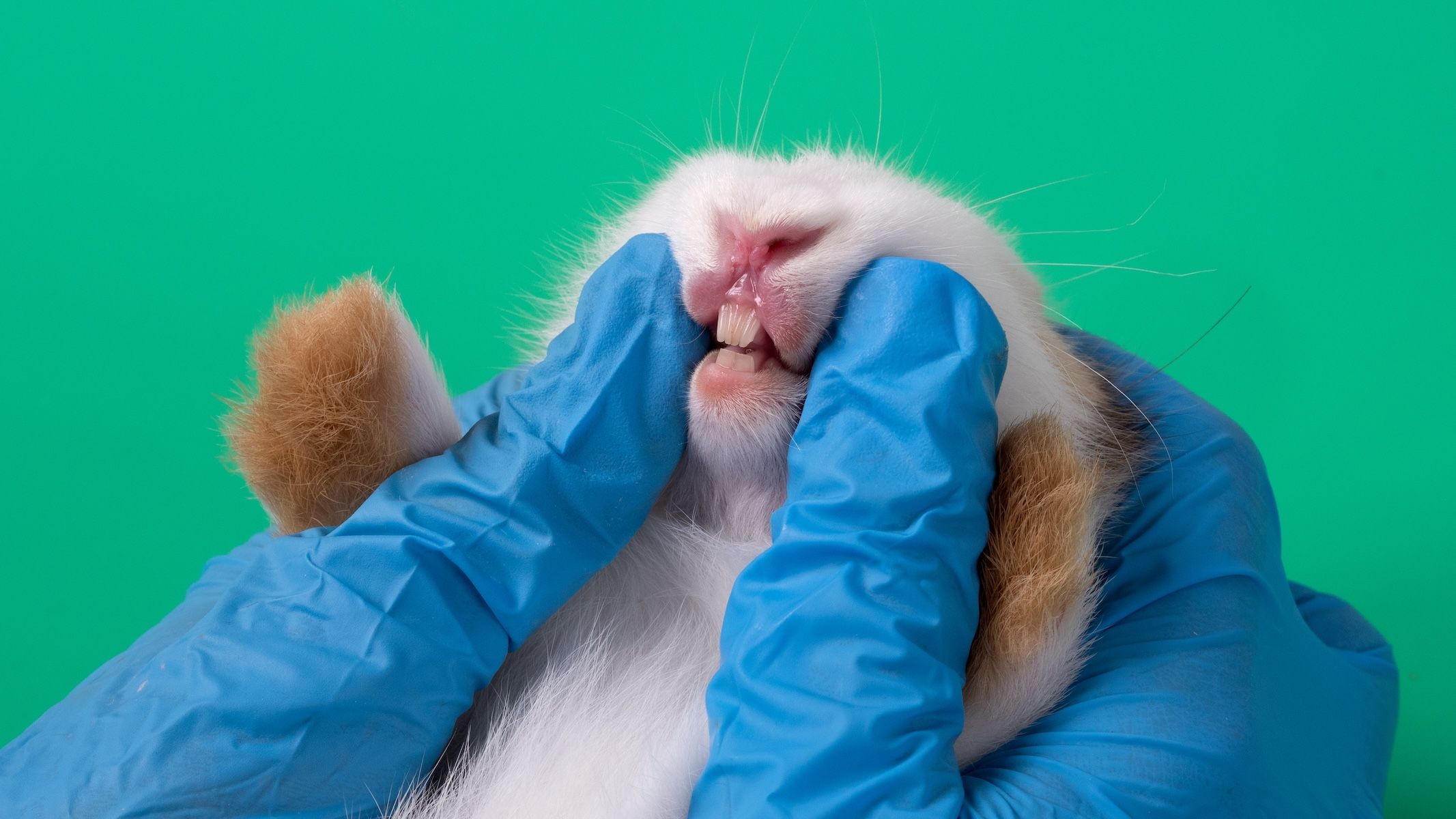
Rabbits have 28 teeth (2 x Incisors 2/1, Canines 0/0, Premolars 3/2, Molars 3/3).2 In the resting jaw, the mandibular incisors are seated behind the central maxillary incisors, in occlusion with the smaller maxillary “peg” teeth just behind them.
Because rabbit jaws are anisognathic—the lower jaw is narrower than the upper—the maxillary and mandibular “cheek” teeth (premolars and molars) never touch.The occlusal surfaces of the maxillary cheek teeth curve buccally, while their mandibular counterparts curve lingually.
Occlusal surfaces of the cheek teeth feature wavy, “zig zag” ridges that create rough surfaces suited to grinding fibrous foods, Proença said. Their side-to-side mastication wears the teeth to a proper trim.
Rabbit teeth are aradicular; they lack true roots. Instead, the elodont—or ever-growing—dentition features open apices lined with germinal cells that continuously spawn fresh, hypsodont (high-crowned) tooth.
If unchecked because of insufficient amounts of nutritional fiber and gnawing materials, these enthusiastic teeth can reach new heights, said Proença. “They can even grow up into the bone.”
Osteolysis and subsequent bone infections may follow, and associated soft tissue often sustains trauma and abscessation.
The right tools
Dentistry in rabbits requires dental tools that conform to the unique architecture of their oral cavities. “Not only is the mouth small,” Proença explained, “but it is also long and dark. So you’re going to need equipment that you don’t use for dogs and cats.”
Although a nasal speculum might aid in evaluating the oral cavity on initial exam, stomatoscopy enables superior visualization of the tunneling bunny mouth to better spot pathologic teeth and fracture lines, and inspect abscess cavities and extraction sites.
It requires a rigid endoscope (2.7mm/18cm/30 degree) with protection sheath, camera and light source. A full set can be purchased online for under $1000, Proença said.
A small mammal dental table is ideal for dental procedures on rabbits, but a well-designed homemade setup can work if it firmly supports the head and permits access for mouth gags, camera and instruments.
Cheek dilators are essential for opening the oral cavity and creating a working corridor. Proença recommended long, slender cheek dilators that can reach the cheek teeth seated caudally, rather than the short, bulky dilators included in generic “rabbit dental kits.”
Molar luxators, approaching 90 degrees in bend and kept sharpened, are critical for breaking down periodontal ligaments tethering cheek teeth.
Supplies should also include fine, curved hemostats or molar extractors bent nearly 90 degrees; these are superior to the chunky extractors in standard kits.
Spatulas should be stocked for retracting soft tissues like tongue and cheek, thereby protecting them from sharp working instruments.
Diagnostic techniques
Initial assessment of the oral cavity might begin with an exam using a nasal speculum. Stomatoscopy can then be deployed to inspect the occlusal surfaces and gingival beds of flagged teeth.
The magnification features of the camera, which can enlarge features as much as tenfold, must be kept in perspective, Proença warned. For instance, the normal ridges and depressions on the occlusal surface of cheek teeth can masquerade as sharp-edged spurs associated with tooth overgrowth and inadequate dietary fiber.
“You see how easily these can look like spurs when you magnify them,” she said. The presence or absence of spur lesions on the buccal and lingual soft tissues can clarify the situation.
The next diagnostic step is imaging, preferably computed tomography (CT). If CT is unavailable, skull radiographs may suffice. Proença recommended heavy sedation to achieve desired positioning. At minimum, she recommended left/right lateral (to include the tympanic bullae) and oblique views, and a ventrodorsal view.
Extractions
Proença advised planning extractions based on thorough review of imaging:Identify diseased or apically elongated teeth, and those associated with abscesses. Decide which teeth to extract, and which to merely adjust.
Next, premedicate the rabbit, and tether it to the table. Then secure the airway through intubation (generally a 2- or 2.5-mm endotracheal tube). These procedures are long and bloody, involve flushing, and carry high risk for aspiration. Proença recommended isoflurane anesthesia, along with the same monitoring parameters used for dogs and cat, like ECG, blood pressure and pulse oximetry.
The veterinarian should then position the table to a comfortable working height and angle. The scope should be held in one’s nondominant hand, saving the dominant hand for manipulating instruments.
Diseased teeth are friable. A tempered approach to extractions— Proença recommended allotting 30 minutes per cheek tooth—may minimize the risk ofshatter. “The name of this pursuit is called patience,” she said. “You will be tempted to put your forceps in, grab the tooth and pull it.But it needs to be so loose that by barely touching it, you can just lift it out.”
Using a molar luxator, work circumferentially to sever dental ligaments, and thenemploy narrow, curved graspers to gently rotate and elevate the tooth. Avoid excessive force, which could crack the crown.
Scope sockets and abscess cavities frequently to reveal hidden fragments and debris requiring removal. Mosquito hemostats work best for fishing out tiny, lodged pieces, Proença advised, but be ready to “dig and dig and dig.” Flush sockets and tracts well, and reinspect before caulking them with sterile packing gel to achieve hemostasis.
The final crucial step is postextraction radiography to confirm removal of all tooth parts. And be prepared to clear away retained shards.
“I cannot tell you how sorry I am for my patients before I started taking x-rays,” Proença said. “I‘d feel certain I’d gotten everything, but x-rays would later show remnants.”
When things go awry
According to Proença, tooth fragment retention is a looming risk for any bunny undergoing extractions. The fallout is infection, inflammation, recurrent abscesses and non-healing tracts.
She shared an old case in which she jiggled a tooth she assessed as loose enough to easily pluck out. The rabbit spooked, and the tooth fractured. To track down the entrapped tooth segment, she widened the socket to the point that the chip was able to migrate out into the sinus and create more havoc.
Rather than “nibbling” repeatedly at one of these dental disasters, she endorses early specialist referral. She also advised referring when one is not equipped for—or experienced with—rabbit dentistry.
Proença’s last word of caution for veterinarians working with rabbits was to refrain from taking initial exam findings at face value. Anesthesia and imaging go a long way in identifying concealed issues within the oral cavity: “Not seeing something in an awake rabbit does not rule out a problem. And sometimes they don’t even have clinical signs,” she said.
References
- Proença L. The art of the pull: mastering rabbit cheek teeth extractions. Presented at: Fetch dvm360 Conference; Long Beach, California. December 5-6, 2025.
- Capello V, Gracis M.In: Lennox AM, ed. Rabbit and Rodent Dentistry Handbook. Ft. Worth: Zoological Education Network, 2005.