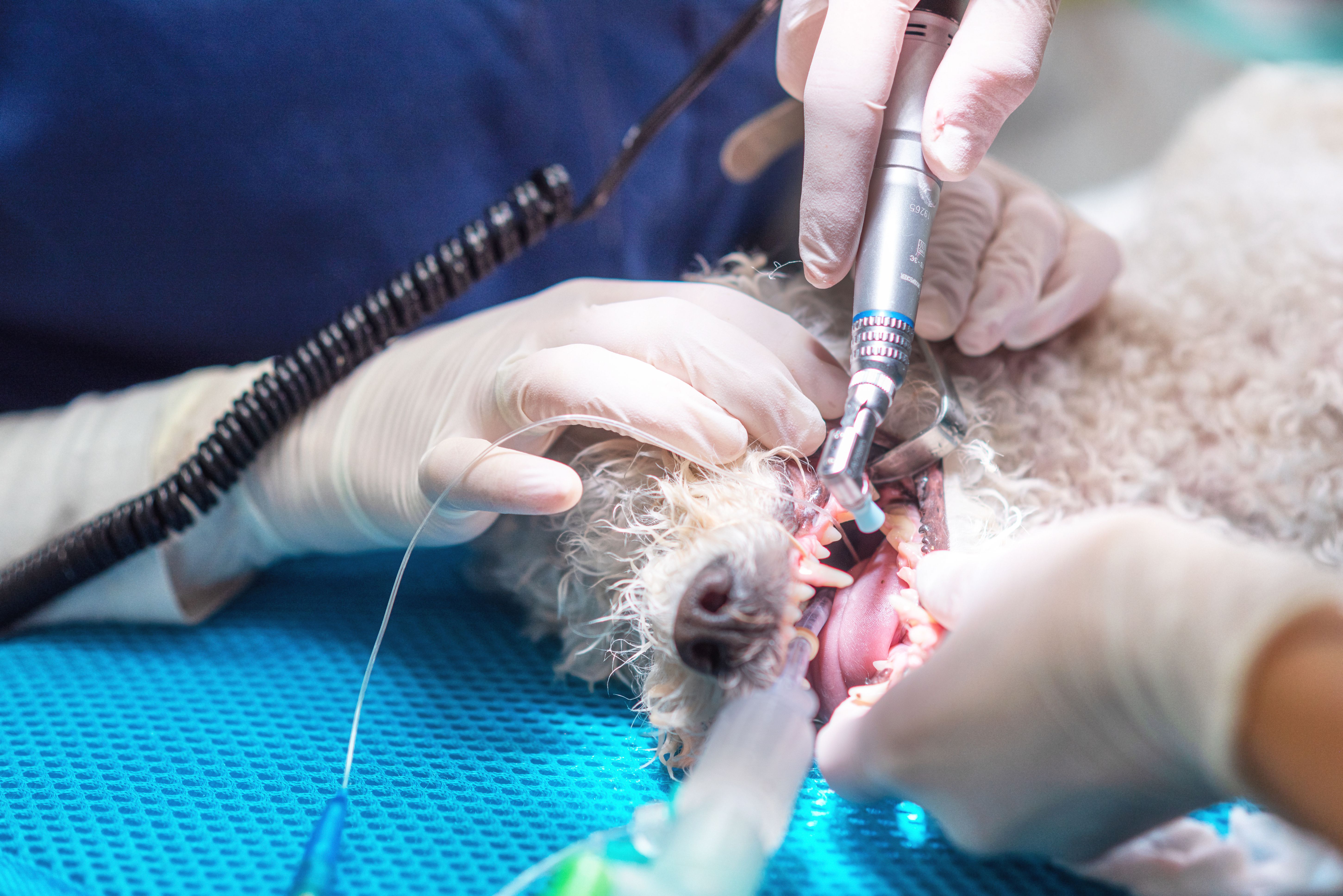
Bite-sized pearls: Practical anesthesia tips for the dental patient
Administration methods and common complications are presented along with barriers and solutions for access to care.
Comprehensive oral health assessment and treatment are widely recognized as a cornerstone of veterinary patient care, recommended annually for companion animals. Dental health in pets is not merely cosmetic; it is intimately linked to systemic health, with untreated periodontal disease contributing to conditions such as cardiovascular disease, diabetes, kidney disease, and respiratory disorders.1
The significance of periodontal disease is often subclinical and undetectable on routine oral exams until it has progressed significantly and silently, increasing the amount of dental work needed, the patient’s time under anesthesia, and thus the degree of pain and the patient’s anesthetic risk. Furthermore, the common practice of deferring dental intervention often results in a patient population that is older with comorbidities, necessitating careful anesthetic planning and more diligent perioperative management.
Barriers and solutions to providing routine veterinary dental care
Primary factors that limit routine dental care in veterinary patients include client fears of anesthesia, especially in the older and medically compromised patient, the veterinary team's lack of confidence in treating higher-risk patients, financial apprehension among veterinary staff, which often leads to subjective assumptions regarding a client’s willingness or ability to invest in recommended care, and genuine financial constraints.
Education and transparent communication are critical to overcoming these barriers. Clients are more likely to consent to recommended procedures when they trust the veterinary team and understand the rationale behind anesthesia and dental care. A 2006 study indicates that veterinary compliance is primarily driven by the strength of the human-animal bond and effective communication rather than client income or service costs. Given that most clients decline recommendations because of a perceived lack of necessity from their provider, rather than financial constraints, comprehensive clinical education is the primary driver of treatment compliance. By strengthening the client-veterinarian bond through better communication, practices can increase compliance by as much as 40%, particularly among underserved populations such as feline patients.2.
Additionally, client anxiety surrounding anesthesia can be minimized by educating clients about your clinic's advanced monitoring devices, development of individualized protocols, and low anesthesia-related mortality rates (0.1%-0.7% for American Society of Anesthesiologists [ASA] class I-II patients; 1%-4% for ASA class III-IV patients).3,4 This may require additional, focused team training in anesthesia and/or the hiring of a board-certified anesthesiologist to provide consultation or direct patient monitoring during the procedure.
Patient assessment and diagnostics
A comprehensive history and physical exam are the cornerstones for appropriate and safe anesthetic planning. The identification of comorbidities (cardiac, renal, hepatic, endocrine) and review of medications inform protocol selection. Annual and preanesthetic bloodwork screen for early detection and subclinical disease, guiding risk assessment and legal defense in case of adverse outcomes. Thoracic radiographs, electrocardiograms (ECGs), +/- a cardiac ultrasound should be recommended for patients with cardiac murmurs, abnormal cardiac rhythm/pulse deficits, or known or suspected pulmonary disease, as well as for brachycephalic patients.
Preanesthetic preparation
Adjustments to chronic medications (eg, withholding antihypertensives or anticoagulants), previsit anxiolytics, and gastrointestinal prophylaxis may be necessary. Protocols such as the chill protocol (gabapentin, melatonin, acepromazine) reduce stress in anxious dogs; gabapentin or trazodone is often used for cats.5 Brachycephalic breeds require special consideration due to potential airway, gastrointestinal, and aspiration risks.6
Preoxygenation before induction increases oxygen reserves and improves safety during apnea.7,8 Initial monitoring (ECG, NIBP) establishes baseline vitals. Anesthetic protocols should be individualized, taking into account the patient’s age, breed, comorbidities, and the type of procedure. A multimodal approach with minimal cardiovascular and respiratory depression is preferred, aiming for anxiolysis, analgesia, muscle relaxation, and immobility.
Anesthetic monitoring
Guidelines for small animal anesthetic monitoring have been established by the American College of Veterinary Anesthesia and Analgesia for 2025.9 It is recommended that the following key vital signs be monitored and documented on a monitoring form every 5 minutes. Key recommendations are as follows:
- Dedicated person to monitor patient/anesthesia
- Digital rectal or esophageal temperature monitoring (every 15 minutes)
- Blood pressure monitoring, ECG, and capnography
- Assessment of mm color, ventilation effort, and SpO2
Common anesthetic complications
- Duration of anesthesia
- Prolonged anesthesia (lasting more than 2 to 3 hours) increases the risk of potential complications.3,4. Cases requiring more extensive intervention should be staged on a case-by-case basis.
- Hypothermia
- May cause dysrhythmias, myocardial depressive effects, reduced hepatic metabolism, coagulopathies, prolonged recoveries, and more. Prevention includes proactive prewarming, active warming devices, and maintaining appropriate room temperatures.
- Hypotension
- SAP greater than 90 mmHg and MAP greater than 60 to 70 mmHg are recommended. Multimodal drug protocols and individualized fluid therapy help minimize risk; inotropes and vasopressors may be required for refractory cases.
- Tracheal trauma
- Dental procedures carry a higher risk of tracheal injury due to frequent repositioning, overinflation of the endotracheal tube (ET), and ET manipulation.10
- Ophthalmic complications
- Ocular trauma is a potential risk during dental procedures for a multitude of reasons. Additionally, tear production decreases significantly under anesthesia; thus, conscious protection of the cornea and lubrication (every 30 minutes), avoiding contact with the cornea, is recommended.11,12
Pain management
Effective pain management is crucial for recovery and quality of life. Dentistry often involves both acute and chronic pain, requiring a multimodal approach.
- Opioids
- Pure μ-opioids (methadone, morphine, hydromorphone) provide profound analgesia and are preferred for extensive dental procedures. Partial agonists (buprenorphine) are suitable for moderate pain. Butorphanol offers sedation but minimal analgesia and is not recommended for dental surgery.
- NSAIDs
- Nonsteroidal anti-inflammatory drugs (NSAIDs) are mainstays for inflammatory pain in dogs and cats, but their usage should be based on individual needs and comorbidities. The author recommends the use of perioperative IV fluid therapy and NSAIDs to be given in the recovery period after the patient has been hydrated and deemed to be normotensive. Galliprant offers targeted anti-inflammatory effects on the EP4 receptor, with fewer potential adverse effects. It may be of benefit to dogs when traditional NSAIDs cannot be used.
- Local and regional anesthetics
- Locoregional blocks provide profound pain control and reduce the need for inhalants. Local anesthetics block nerve impulse transmission and nociception and should be used when dental extractions or oral surgery will be performed, and there are no other contraindications (eg, neoplasia, coagulopathies, etc).
Recovery
One critical morbidity and mortality study showed that postoperative deaths accounted for 47% of anesthetic-related deaths in dogs and 61% in cats, and nearly half of those occurred in the first 3 hours after extubation.3 Close monitoring and maintenance of normothermia are crucial in the postoperative period.
Amber Hopkins, DVM, cVMA, CCRT, DACVAA, serves as vice president of medical operations for Thrive Pet Healthcare, as well as national specialty director of anesthesia and analgesia and chair of the National Specialty Directors. She is board-certified in veterinary anesthesia and analgesia and holds advanced certifications in veterinary medical acupuncture, canine rehabilitation, pain management, and extracorporeal therapies.
References
- Lohiya DV, Mehendale AM, Lohiya DV, Lahoti HS, Agrawal VN. Effects of periodontitis on major organ systems. Cureus. 2023;15(9):e46299. doi:10.7759.cureus.46299
- Lue TW, Pantenburg DP, Crawford PM. Impact of the owner-pet and client-veterinarian bond on the care that pets receive. J Am Vet Med Assoc. 2008;232(4):531-540. doi:10.2460/javma.232.4.531
- Brodbelt DC, Blissitt KJ, Hammond RA, etc. The risk of death: the confidential enquiry into perioperative small animal fatalities. Vet Anaesth Analg. 2008;35(5):365-373. doi:10.1111/j.1467-2995.2008.00397.x
- Redondo JI, Otero PE, Martínez-Taboada F, Doménech L, Hernández-Magaña EZ, Viscasillas J. Anaesthetic mortality in dogs: a worldwide analysis and risk assessment. Vet Rec. 2024;195(1):e3604. doi:10.1002/vetr.3604
- Costa RS, Jones T, Robbins S, Stein A, Borns-Weil S. Gabapentin, melatonin, and acepromazine combination prior to hospital visits decreased stress scores in aggressive and anxious dogs in a prospective clinical trial. J Am Vet Med Assoc. 2023;261(11):1660-1665. doi:10.2460/javma.23.02.0067
- Downing F, Gibson S. Anaesthesia of brachycephalic dogs. J Small Anim Pract. 2018;59(12):725-733. doi:10.1111/jsap.12948
- McNally EM, Robertson SA, Pablo LS. Comparison of time to desaturation between preoxygenated and nonpreoxygenated dogs following sedation with acepromazine maleate and morphine and induction of anesthesia with propofol. Am J Vet Res. 2009;70(11):1333-1338. doi:10.2460/ajvr.70.11.1333
- Ambros B, Carrozzo MV, Jones T. Desaturation times between dogs preoxygenated via face mask or flow-by technique before induction of anesthesia. Vet Anaesth Analg. 2018;45(4):452-458. doi:10.1016/j.vaa.2018.03.004
- Bailey K, Briley J, Duffee L, et al. The American College of Veterinary Anesthesia and Analgesia Small Animal Anesthesia and Sedation Monitoring Guidelines 2025. Vet Anaesth Analg. 2025;52(4):377–385. doi:10.1016/j.vaa.2025.03.015
- Mitchell SL, McCarthy R, Rudloff E, Pernell RT. Tracheal rupture associated with intubation in cats: 20 cases (1996-1998). J Am Vet Med Assoc. 2000;216(10):1592-1595. doi:10.2460/javma.2000.216.1592
- Raušer P, Novák L, Mrázová M. Influence of anaesthetics on aqueous tear production in dogs: a systematic review. Vet Anaesth Analg. 2022;49(6):525-535. doi:10.1016/j.vaa.2022.08.007
- Bedos L, Allbaugh RA, Roy M, Kubai MA, Sebbag L. Precorneal retention time of ocular lubricants measured with fluorophotometry in healthy dogs. Vet Ophthalmol. 2023;26(suppl 1):81-88. doi:10.1111/vop.13065